Molar pregnancies are fairly rare, happening with roughly 1 case for every 600 pregnancies in the UK. When a molar pregnancy arises a problem occurs at the time of conception, when the egg and sperm join together that results in the formation of cells that grow very rapidly but are unable to form the placenta and foetus of a normal pregnancy.
Molar pregnancies take two different forms, complete and partial molar pregnancies. These differ in their genetic make-up, their development and particularly in the risk of needing additional treatment.
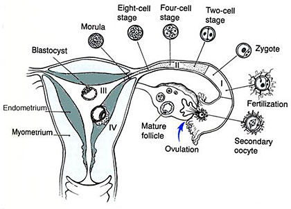
Figure 1. Normal Egg Development
The diagram in Figure 1 is a reminder of how an egg normally develops, is fertilised and then implants in the wall of the uterus. In a molar pregnancy the steps are identical except that at the time of fertilisation there is a problem with either the maternal chromosomes being lost as in a complete mole or there being two sets of chromosomes from the father and one from the mother as in a partial molar pregnancy.
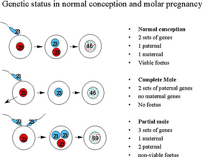
Figure 2.
The diagram in Figure 2 shows the genetics of how the two different types of molar pregnancy arise and some of their important features.
In a complete molar pregnancy the genetic material is just from the father as the original nucleus containing the mother’s genetic material is lost at the time of conception or whilst the egg is developing in the ovary. Complete molar pregnancies form a mass of rapidly growing cells but do not contain a foetus and can not develop into a baby. After diagnosis and evacuation there is about a 10-15% chance of needing further treatment.
In a partial molar pregnancy there is genetic material from both the father and the mother but an imbalance as there two sets from the father. In a partial molar pregnancy there can be a foetus visible on an early ultrasound, but it is always abnormal and cannot survive. After the evacuation, most partial molar pregnancies do not require any additional treatment as in more than 99% any of the residual cells just die away over the following weeks.
In the first few weeks of a molar pregnancy there is often a tendency for morning sickness, bleeding and some abdominal pain. However these symptoms do not always occur and can also occur in a normal pregnancy. Most complete molar pregnancies are diagnosed at the first ultrasound scan. In a complete molar pregnancy the scan shows a mass of cells without the presence of a foetus. In a partial mole an abnormal non-viable foetus and placenta may be seen.
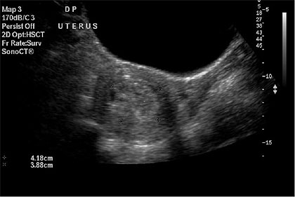
Figure 3. An ultrasound showing a complete molar pregnancy before evacuation.